Home / Departments / Radiology & Nuclear Medicine Services
ClearMedi Healthcare provides high end Radiology and Nuclear Medicine services to various Hospitals in India besides running these departments at it’s own/ managed hospitals and Cancer Centres. A team of over 50 highly skilled and experienced Radiologists and Nuclear Medicine Specialists work with best-in-class machines and equipment to deliver quality radiology services.
- Clearmedi Radiant Hospital Mysore
- Clearmedi Hospital & Cancer Care Hospital, Vasundhara
- Shanti Mukand Hospital and Cancer Care Centre, Delhi
- Hamdard Imaging Centre at Jamia Millia University, Delhi
- Paras Hospitals Gurgaon
- Paras HMRI Hospital Patna
- Paras Hospitals Panchkula
- Paras Global Hospital Darbhanga
- Paras Bliss Hospital Panchkula
The group operates radiology and nuclear medicine equipment at various hospitals above, as below:
- Two 3T MRI scanners
- Four 1.5T MRI scanners
- Three 128 slice CT scanners
- Four 16 slice CT scanners
- Three advanced PET CT scanners with TOF technology
- Multiple advanced ultrasound systems
- Multiple units of fully digital X Ray systems as well as CR systems
- Five Bone densitometry Units
- Five Mammography Units
A team of over 50 highly skilled and experienced Radiologists and Nuclear Medicine Specialists work with best-in-class machines and equipment to deliver quality radiology services. The Radiologists and Technologists working atClearMedi are highly skilled with sub-specialty experience. All the nine radiology departments at various sites across India are linked on an advanced PACS system facilitating cross reporting. The team works in cohesion with clinicians to provide a superlative degree of medical care to patients. They are also compassionate and empathetic in their approach. They take utmost care to build a comfortable and stress-free environment at the facility. Our staff works hard to ensure that patients do not feel rushed at any point in time. The centres are accredited by NABH.
The group attaches great importance to quality imaging and academic research. Training programs, CMEs and other activities are regularly held under the banner of Clearmedi Healthcare to further learning. Hamdard Imaging Centre is recognized by MCI to provide undergraduate teaching to students at Jamia Hamdard University. The Radiology Department at Paras Hospitals, Gurgaon is accredited by National Board of Examinations to impart post graduate training (DNB).
Radiology is the branch of medicine that uses medical imaging and X Rays to diagnose and treat diseases/injuries. The specialty is bed rock of disease diagnosis and management in current times. It is vital for management of patients in all specialties. With expert execution and interpretation, the imaging can help in early diagnosis, accurate staging and evaluation of the disease status. Radiology and Imaging includes various modalities, as below:
- Radiography: X-rays are a form of electromagnetic radiation used to get a radiographic image of bones, organs and tissues.
- CT Scan: Computed Tomography is a technique that uses computer-processed images obtainedwith X-rays from various angles. This technique allows the experts to see inside an organ without actually cutting it. CT scan is useful for imaging of head, neck, lungs, angiography and cardiac anatomy.
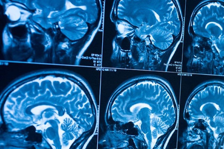
- MRI: Magnetic Resonance Imaging is a radiation free modality which uses uses magnetic fields and radio waves for imaging. MRI can give pictures of the anatomy and physiological processes of the body. It has wide use in all parts of the body, more so in brain and abdomen.
- Ultrasound: It is a modality which uses sound waves to produce real-time images. It is radiation free and completely safe. It is commonly used for diagnosis of abdominal diseases, fetal imaging and in musculoskeletal system.
- Mammograms Low energy X-rays are used to diagnose and screen breast cancer.
- Fluoroscopy: X rays are used to produce real-time moving images of the body. It is particularly useful for carrying out medical procedures such as inserting stents and drainage catheters.
- Nuclear medicine: It is a sophisticated procedure of using radioactive substances for diagnosis and treatment of diseases. It uses equipment like Gamma Camera and PET-CT scanner.
- Interventional Radiology: Images are used to perform minimally invasive procedures. They are specifically useful in treating and diagnosing patients without open surgery.
ClearMedi is committed to providing exceptional quality of treatment to the patients and Radiology is an important constituentto achieving that. The department provides a wide spectrum of services that range from the most basic imaging to the most complicated interventions. Here are examples of services that ClearMedi Radiology services provides.
- Advanced Neuroimaging
- Abdominal Imaging & Intervention
- Breast Imaging
- Cardiovascular Imaging
- Emergency Imaging
- Musculoskeletal Imaging
- Neurological Imaging
- Neurological Intervention
- Nuclear Medicine and Molecular Imaging
- Pediatric Imaging
- Thoracic Imaging & Intervention
- Vascular Imaging & Intervention
Nuclear Medicine is a specialized medical discipline that uses radioactive tracers or radioisotopes to obtain images of the body. In effect, it is the reverse of radiology as it measures the radiation emitting from the body. These procedures are safe, painless and effective in diagnosis and treatment. The radiotracers are given either intravenously, orally or through inhalation. These tracers enable a very sensitive technique to detect abnormalities in an organ. It gets concentrated in the problem area and gives an accurate vision to the specialists. The radiopharmaceuticals emit radiation that travel only a short distance
Nuclear medicine uses the following imaging techniques
- PET CT Scan – Positron emission tomography scans are one of the most advanced levels of imaging. PET scans produce three-dimensional images of the organs and tissues. PET is particularly useful in detecting cancer earlier than other imaging techniques. It has a unique way of detecting the changes in the biochemistry of the body that occurs before the formation of a malignant tumor. It is also helpful in evaluating the effects of cancer treatments.
- Gamma Camera – Also known as Scintillation Camera, it is an effective and powerful diagnostic device. The gamma camera is variable-angle diagnostic equipment. A small amount of radioactive pharmaceutical is given to the patient who is then asked to lie on a flat table. The pharmaceutical depends upon the specific organ or tissue that is under examination(such as heart, lungs, bone, kidney, liver, gall bladder function, thyroid and brain). The amount given to the patient is insignificant and doesn’t last a very long time. It is an effective and painless way to detect the spread of the disease.
